")
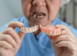
In modern dentistry, X-rays are considered an indispensable tool for diagnosing many conditions that cannot be seen with the naked eye. By using electromagnetic waves, X-rays provide precise images that help dentists evaluate the health of teeth and the surrounding tissues, which contributes to developing effective and appropriate treatment plans for each case.
The importance of X-rays in dentistry
Early detection of hidden problems:
X-rays are used to detect tooth decay in its early stages, especially in areas that are difficult to see, such as between teeth or under fillings. They also help detect root infections and abscesses that may develop without clear symptoms.
Accurate planning of dental treatments:
X-rays provide precise information about bone structure and density, which helps in planning treatments such as orthodontics or dental implants. Images are used to determine optimal implant placement and assess the need for additional procedures.
Monitoring children’s tooth development:
In pediatric dentistry, X-rays are used to monitor tooth growth and development and to ensure there are no impacted teeth or problems in the transition from primary to permanent teeth.
Detection of early jaw tumors:
X-rays can detect changes in the jawbone that may indicate the presence of tumors or cysts, allowing for early diagnosis and immediate treatment.
How does the X-ray device work?
A small amount of ionizing radiation is passed through the body. In the past, this was placed on a sheet of special film. Nowadays, X-ray examinations are more likely to use a device that captures the transmitted X-rays to create a digital image.
Calcium in bones and teeth prevents radiation from passing through, so healthy bones and teeth appear white or gray. On the other hand, radiation passes easily through other tissues except teeth and bones, so they do not appear clearly on the X-ray.
Latest types of dental X-rays
Dental X-rays vary according to their purpose, whether for early detection, treatment planning, or post-treatment follow-up. They are generally classified into two main types: intraoral and extraoral X-rays. Below is a detailed explanation of each type:
Intraoral X-rays:
These are the most common in dental clinics, where films or digital sensors are placed inside the patient’s mouth to capture images of the teeth and supporting tissues from different angles.
1- Periapical X-ray
Used to image one complete tooth, from the crown to the root and surrounding bone.
It helps in diagnosing:
- Tooth root infections
- Abscesses or cysts around the root
- Microfractures in the tooth or root
- The success of root canal treatment
- It is essential before dental implant placement or extraction of affected teeth
2- Bitewing X-ray
Used to capture upper and lower posterior teeth in one image. It focuses on the spaces between teeth where decay is difficult to see with the naked eye. It is useful for:
- Early detection of interproximal caries
- Monitoring the condition of dental fillings and ensuring no leakage beneath them
- Evaluating bone density around teeth in periodontal disease cases
3- Occlusal X-ray
Shows a full arch of teeth in either the upper or lower jaw from an occlusal perspective. It is used to detect:
- Supernumerary or impacted teeth
- Foreign objects inside the mouth
- Fractures in the floor of the jaw or the palate
- Tooth development problems in children
Extraoral X-rays:
In this type, the X-ray device is placed outside the mouth and is used to image the entire jaw or skull for broader evaluation purposes.
1. Panoramic X-ray
Shows all teeth, upper and lower jawbones, the temporomandibular joint (TMJ), and the sinuses. It does not show fine details of each tooth but is used for:
- Orthodontic treatment planning
- Evaluating impacted teeth such as wisdom teeth
- Detecting cysts or jaw tumors
- Monitoring jaw fractures after trauma
It is preferred before undergoing major oral surgical procedures.
2. 3D imaging (CBCT – Cone Beam Computed Tomography)
One of the latest and most accurate technologies for imaging the mouth and jaws, providing a detailed three-dimensional image of bones, teeth, nerves, and surrounding tissues. Ideal for:
- Dental implants to determine depth and location with high precision
- Complex surgeries such as removing teeth close to nerves
- Evaluating the temporomandibular joint (TMJ)
- Planning advanced cosmetic treatments
3. Digital radiography
An advanced technology that uses digital sensors instead of traditional films. Its main advantages include:
- Reducing radiation exposure by up to 80%
- Displaying images instantly on a computer screen
- Ability to enlarge images and adjust contrast to clarify fine details
- Easy storage and comparison of images in future visits
It is considered the safest option for pregnant women and children due to lower radiation exposure.
Technologies integrated with X-ray types:
Artificial Intelligence (AI): Used to analyze X-ray images and automatically detect decay and fractures.
Integration with ultrasound imaging: Used in some cases to evaluate soft tissues surrounding the teeth and jaw.
Contrast radiography: May be used with contrast materials such as oral or rectal contrast in diagnosing jaw or digestive-related issues, although it is rarely used in dentistry.
Ultrasound: Uses sound waves to create images of internal body structures.
Magnetic Resonance Imaging (MRI): A combination of magnetic fields and radio waves to produce 3D images of tissues other than bones and teeth, which do not appear very clearly.
Are there risks from dental X-rays?
Yes, but exposure to X-ray radiation is extremely low, making it safe for both children and adults. If your dentist uses digital X-rays instead of traditional film methods, radiation exposure risks are even lower.
This is what we ensure at Wonders Dentistry by using the latest digital radiography technologies to reduce risks for our patients, even if they are already minimal.
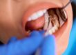
In a dental clinic, when taking images of one or two teeth using an intraoral X-ray device, you will sit in a chair and the X-ray machine will be positioned next to your head to capture images of your mouth. Some dental clinics have a separate X-ray room, while others perform it in the same room.
Radiation safety procedures
- Use of protective shielding: A lead apron is used to protect sensitive body areas such as the thyroid gland and abdomen from unnecessary radiation exposure during imaging.
- Digital imaging techniques: Preferred due to their ability to reduce radiation dose, making them a safer option for patients.
- ALARA principle (As Low As Reasonably Achievable): Applied to ensure the lowest possible radiation dose is used to achieve diagnostic goals while maintaining patient safety.
- Safe time intervals between sessions: The dentist determines appropriate intervals between X-ray sessions based on the patient’s condition and needs to minimize repeated exposure.
Cases that require X-ray imaging
Before surgical tooth extraction:
X-rays are used to evaluate the position of the tooth to be extracted and its relation to surrounding structures, helping plan the procedure and reduce risks.
When impacted teeth are suspected:
X-rays help determine the position of impacted or extra teeth and assess their effect on adjacent teeth and bone.
Diagnosis of advanced periodontal disease:
X-rays are used to evaluate bone loss around teeth, helping determine disease severity and plan treatment.
After jaw and facial injuries:
X-rays are used to assess damage to bones and teeth after trauma and determine the need for medical or surgical intervention.
After dental X-ray imaging
When images are immediately available, as in digital X-rays, your dentist will review them and check for any abnormalities. If an endodontist is cleaning your teeth after an infection, for example, the dentist may review the X-ray results with you after the procedure.
If your dentist finds problems, such as tooth decay or any surgical issues, they will discuss your treatment options. If no problems are found, continue maintaining good oral health habits.
Conclusion
Dental X-rays play a vital role in accurate diagnosis and treatment planning in dentistry. When used safely and appropriately, they help detect many problems at early stages, contributing to better patient care. If you experience any unusual symptoms or wish to assess your oral health, do not hesitate to consult your dentist, who is best qualified to determine the need for X-rays and guide you toward optimal treatment.
FAQs
Why do I need X-rays if I don’t feel any pain?
Pain is often the last sign of a problem. X-rays, especially bitewing X-rays, allow us to see interproximal decay (hidden between teeth) and early bone loss that cannot be seen with the naked eye. Detecting these issues early means simple fillings instead of root canal treatment or extraction later.
Is it true that the “lead apron” is no longer necessary during imaging?
Yes, this is the modern global standard. Recent studies have shown that advanced digital X-ray devices provide highly precise beams with extremely low doses, making the lead apron medically unnecessary and sometimes even interfering with image clarity. However, we always provide it for patients who feel more comfortable using it.
What is the difference between panoramic X-rays and 3D CBCT scans?
Panoramic X-rays provide a flat (two-dimensional) image of the entire jaw and are excellent for evaluating wisdom teeth and overall bone health. CBCT scans create a three-dimensional model of teeth and nerves. They are used for precise procedures such as dental implant planning to avoid vital nerves and ensure 100% accuracy.
Are dental X-rays safe during pregnancy?
Yes, diagnostic X-rays are considered safe when medically necessary. The radiation dose from digital X-rays is extremely low and directed only at the mouth, not reaching the abdominal area. Medically, leaving an abscess or dental infection untreated poses a greater risk to the fetus than the X-ray itself. We use the fastest digital sensors to minimize exposure time as much as possible.